 #EuropeanRadiologyCares
Cranium florens
An art installation to raise awareness of the significance of radiology and its role in modern medicine and healthcare, particularly for the benefit of patients.
Cranium florens is an artwork created by Pablo Spitzer, designer and custom artist, and commissioned by the European Society of Radiology.
It was inspired by ECR 2017 President, Prof. Dr. Paul Parizel, who has always loved the imagery of skulls with flowers as two symbols combining Death and Life/Love.
What is radiology?
Radiology, also called diagnostic imaging, is a series of different tests that take pictures or images of various parts of the body. These tests are unique in that they allow doctors to visualise the inside of your body.
This is the central role that the field of radiology plays in modern medicine.
With a range of imaging methods at their disposal, the modern doctor can peer into the depths of your body to discover the root cause of your illness.
Medical imaging is essential in the diagnosis of many diseases and conditions, including internal injury, head injury, cancer, broken bones, torn muscles and tendons, heart conditions, and much more.
Radiology involves various healthcare workers who work as a team to handle a patient at various stages and procedures. A typical radiological screening might involve the following individuals:
A physician requests a screening.
A nurse takes care of the patient before and after the screening.
The radiographer or radiological technologist uses the imaging equipment to get the best possible image of the requested area.
The radiologist, a trained medical doctor, interprets the medical image and communicates what they find to the physician, sometimes also to the patient directly. As a member of a team of doctors, the radiologist works with other physicians to achieve the best outcome for the patient.
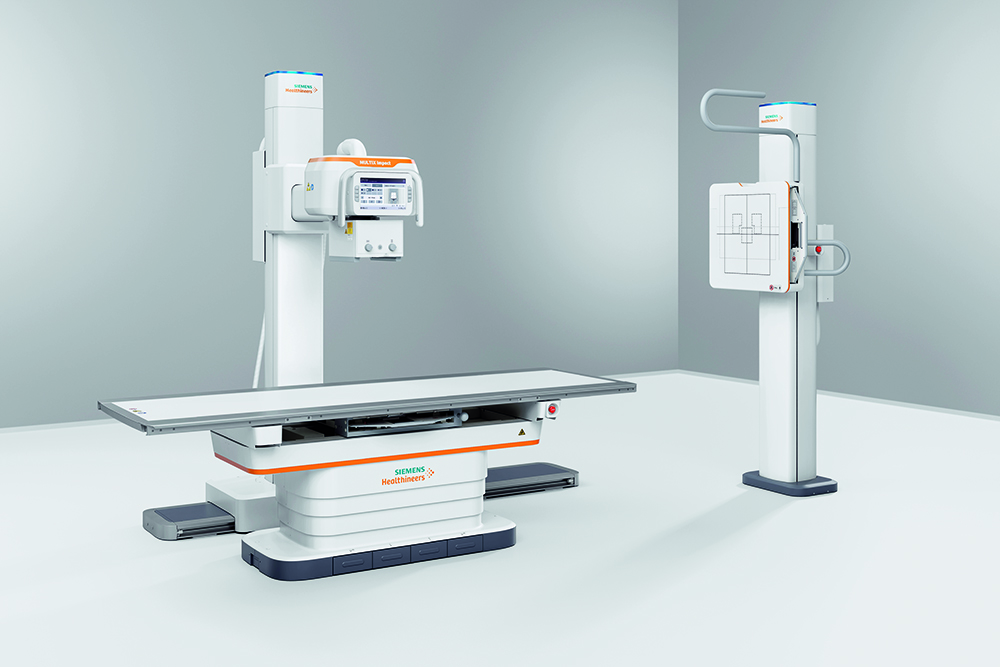
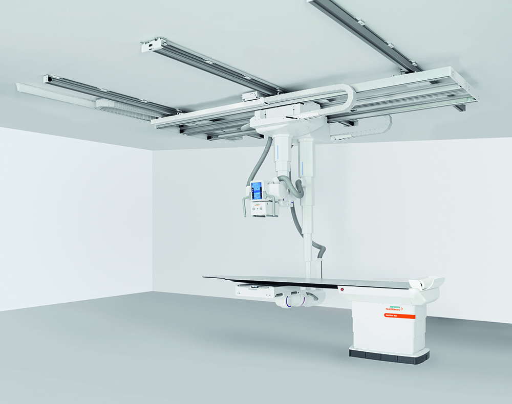
X-Ray Radiography
X-rays are a form of electromagnetic radiation, similar to light but of shorter wavelength and capable of penetrating solids and ionising gases. A radiograph is a photographic image produced by the action of x-rays.
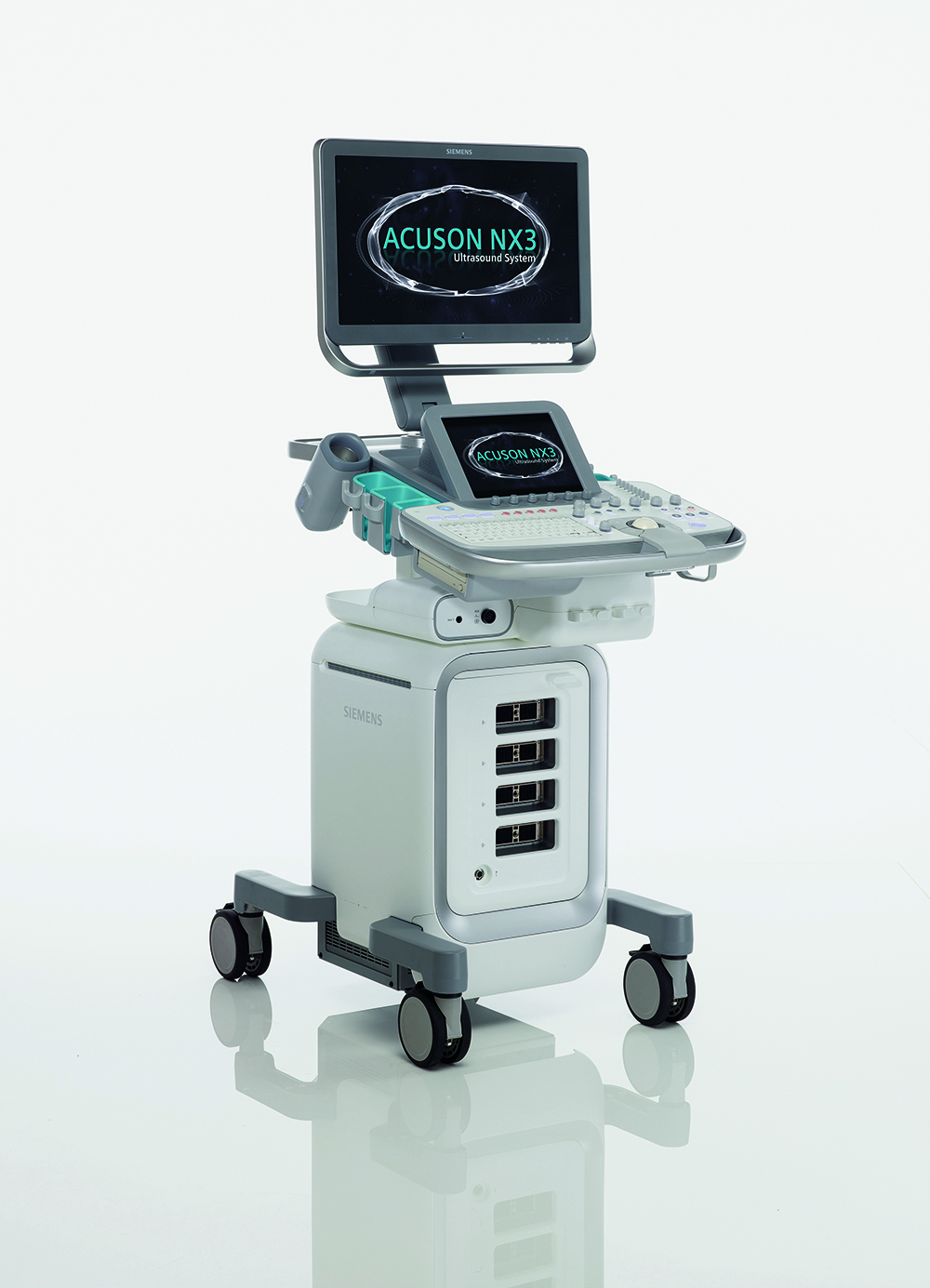
USUltrasound
Ultrasound, also known as sonography, uses high-frequency sound waves to look at organs and structures inside the body. Healthcare professionals use it to view the heart, blood vessels, kidneys, liver, and other organs.
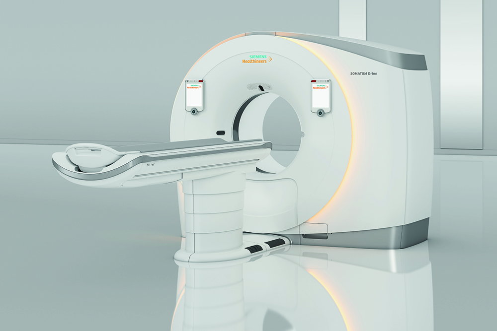
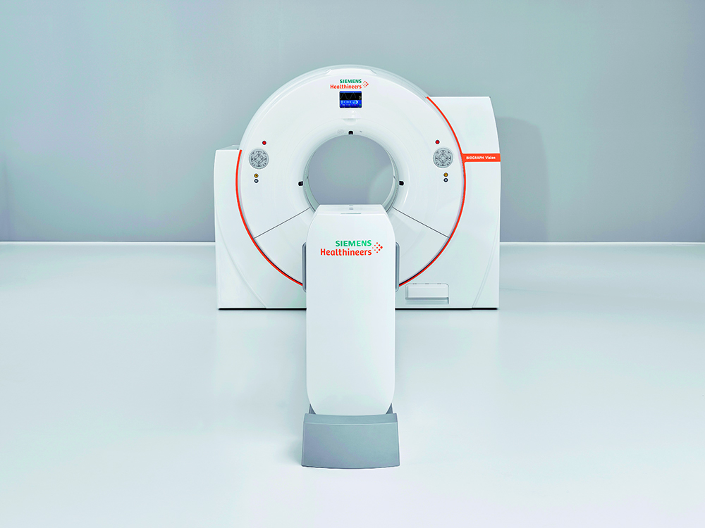
CT Computed tomography
In computed tomography (CT), a computer controls the motion of the x-ray source and detectors, processes the data, and produces the image. Tomography refers to a technique for displaying a representation of a cross-section through a human body or other solid object using x-rays or ultrasound.
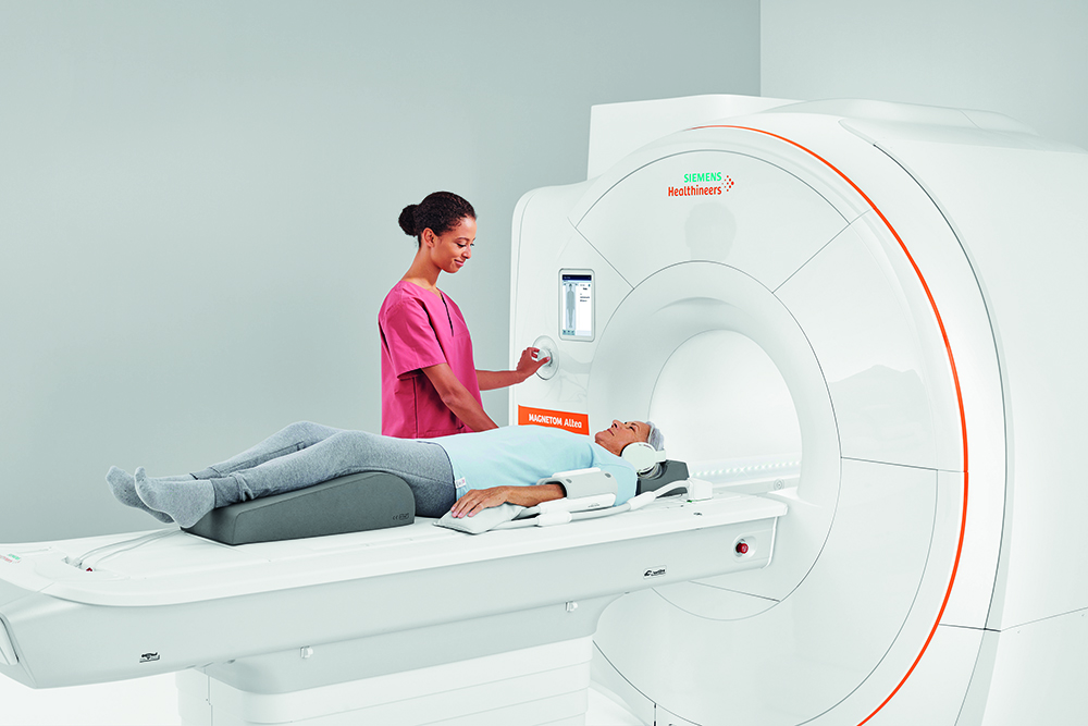
MRI Magnetic resonance imaging
Magnetic resonance imaging (MRI) uses strong magnetic fields, magnetic field gradients, and radio waves to generate images of the organs in the body. MRI machines do not use ionising radiation and as a result, carry no risk of even minute damage to DNA or cellular matter. MRI scanners are particularly well suited to image the non-bony parts or soft tissues of the body.
PET Positron emission tomography
Positron emission tomography (PET) is an imaging technology in which substances containing positron-emitting isotopes are introduced into the body, allowing the precise location of physiological processes by detection of the gamma rays produced by the isotopes.
Fluoroscopy
Fluoroscopy is an imaging technique that uses x-rays beams to obtain real-time moving images of the interior of an object. The beam is transmitted to a TV-like monitor so that the body part and its motion can be seen in detail.
All images kindly provided by Siemens Healthineers.
ESR European Society of Radiology
The European Society of Radiology (ESR) is an international medical society based in the centre of Vienna, in the ‘House of European Radiology’.
With more than 120,000 members across the globe, it has grown to become the largest radiological society in the world. The ESR serves as an umbrella organisation for European radiologists (also representing them at EU level), organises the annual European Congress of Radiology (ECR), supports education through various activities of the European School of Radiology (ESOR) and the European Board of Radiology (EBR), and coordinates the publication of European Radiology, a prestigious monthly peer-reviewed medical journal, as well as other journals.
The core of the ESR’s activities is the organisation of its annual meeting, the European Congress of Radiology (ECR). With more than 30,000 participants from around 100 countries, the ECR has developed into the largest radiological meeting in Europe since it first took place in Vienna in September 1991.
The ECR is held annually in Vienna in late February or early March and is officially recognised as a Green Meeting by the Austrian State.
Media proprietor and responsible for the contents:
European Society of Radiology (ESR)
myESR.org
ZVR No.: 083757049
VAT No.: ATU65092507
Headquarters:
Am Gestade 1, 1010 Vienna, Austria
Phone: +43 1 533 40 64-0, Fax: +43 1 533 40 64-448
E-mail: communications@myESR.org
Executive Director: Peter Baierl
Objects of the society are the coordination and organisation of the annual meeting, the European Congress of Radiology, as well as of other congresses, lectures and educational activities, membership management, publication of scientific and professional information, and various e-learning initiatives.
Place of jurisdiction is Vienna, Austria.